


The bowel is part of our digestive system and it works to digest the food we eat, absorb the goodness and nutrients into our blood stream, then process and expel the waste that the body cannot use.
The digestive system works by pushing food through the intestines which usually takes between 24 to 72 hours. Muscular contractions squeeze (peristalsis) the food through the different sections of the intestine. These different sections are separated by bands of muscles, or sphincters, which act as valves.
The passage of food from one area of the intestines to another is coordinated so that food stays in a specific area for long enough for the gut to do a particular job – absorb fluids and nutrients, or process and expel waste.

The small bowel (small intestine)
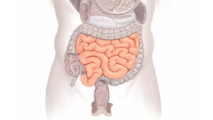
The small bowel (or small intestine) is around 6 – 8 m long and roughly 2cm wide.
There are 3 parts of the small bowel (small intestine): the duodenum, the jejunum and the ileum.
Food passes from the stomach into the duodenum, which is the tube that leads from the stomach into the intestines. The food then passes through the jejunum and ileum before going to the large bowel (colon). The small bowel (small intestine) absorbs nutrients and much of the liquid from foods. At the point where food is passed from the small bowel into the large bowel (colon) it is of a ‘porridge like’ consistency.
The Large Bowel (Colon – large intestine)
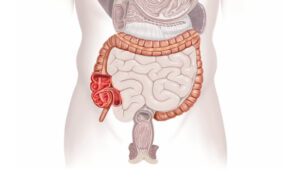
The large bowel (also known as the colon, or large intestine), starts at the final portion of the small bowel (small intestine) and goes all the way to the rectum. The large bowel (colon) is about 2m long and 6-7 cm wide.
This muscular tube is made up of the ascending colon, the transverse colon and the descending colon which ends at the rectum and the anus. The colon’s most important job is to store, process and get rid of waste. The colon also absorbs some nutrients and water. Key to this process are the hundreds maybe thousands of bacteria resident in the colon – both ‘good’ and ‘bad’ – which collectively make up the gut flora. The rectum can store waste but most of the time it’s empty. Waste is expelled through the anus.
The rectum and muscles
Once the bowel has done its work and absorbed nutrients from food, the waste travels to the rectum which stretches, triggering a message to the brain to say that the bowel is full and needs to be emptied. The pelvic floor muscles, when well-toned, ensure the anus remains closed until it’s time to go to the toilet.
The rectum and anus
After food has travelled along the gut, it has become digested and the nutrients and fluids absorbed; the waste is then expelled through the rectum and anus.
The rectum and the upper portion of the anal canal are richly supplied with nerves. When the rectum is full, the nerves sense this fullness and then inform the brain whether this is due to gas or stool.
When we need to go to the toilet, the brain tells the anal sphincter muscles, via the nerves, to relax. As the muscles relax, the anus opens and the rectum empties. In some neurological and spinal conditions the brain cannot tell whether the bowel is full of waste (faeces) or just wind. This can lead to accidental leakage.
Anal sphincters
The anal canal is about 3 – 4cm long in women and sometimes slightly longer in men. The internal and external sphincters form 2 concentric rings which run along the length of the anal canal.
The internal anal sphincter (IAS) is made of smooth muscle and we do not have voluntary control of this muscle. It works automatically to keep the anus closed until we are ready to have a bowel movement.
The external anal sphincter (EAS) is made of striated muscle (the same as the pelvic floor muscles); we do have voluntary control over the EAS – allowing us to hold on if we are aware of wind or diarrhoea.
The pelvic floor muscle
The pelvic floor muscles are layers of muscle stretched like a sheet from the pubic bone in the front, to the bottom of the backbone (coccyx). There are 3 openings through the pelvic floor in women and 2 in men -the anus (back passage), the vagina in women (birth canal) and the urethra (bladder outlet). The muscles support these 3 openings, but if they are weakened or not in good condition they cannot support the openings effectively.
An important part of the pelvic floor muscle regarding bowel control is the deepest layer called Levator Ani – which directly translated from Latin means ‘lift the anus’. One of the muscle groups within levator ani is the puborectalis. This muscle forms a supportive strap around the junction between the rectum and the anal canal and helps to keep us in control of our bowels by forming a flap like valve, which prevents stool passing too easily into the anal canal.
The sacral nerves
The brain tells the bowel what to do by sending electrical signals to the muscles in the pelvic floor, the sphincters and the urethra.
The commands from the brain to the bowel (colon) are sent as electrical impulses and are carried by a system of special fibres called nerves.
These signals start in the brain and go to the spinal cord and continue to the nerves located in the sacral area of the back.
Some of these sacral nerves go to the rectum, levator ani muscle, and external sphincter muscles, controlling their activities.
Two key sacral nerves that are vital to the functioning of the bowel are the pudendal nerve and the pelvic splanchnic nerve.
Through a series of reflexes and signals the nerves in the bowel is coordinated with the pelvic floor muscles and anal sphincters in order to store bowel contents until there is an appropriate place to go to the toilet and then to allow complete bowel emptying once on the toilet.
This coordination ensures that the sphincters remain closed, opening only during defecation. So, when the rectum fills, and the pressure inside it increases, the nerves sense the pressure and tell the brain about it. The brain then sends signals via the nerves to keep the external sphincter closed. Normally, this prevents leakage and is called the guarding reflex.
When you need to go to the toilet, the brain tells the nerves to signal the rectum to empty, and relax the muscles surrounding the anus. The rectum empties and stool is expelled.
Sensory signals
The nerve pathways are also shared with fibres that carry sensory signals, such as pain or fullness. Pain that originates in the pelvic area is transmitted along these pathways to the sacral area, up the spinal cord and back to the brain.
The same happens as the rectum fills. This sensation of filling is registered by the nerves, which transmit the information to the brain.
The nerves can also tell the brain whether it’s gas or stool that’s filling the rectum.
For the bowel to function and work properly, and to relieve constipation, you need:
- The nerves of the rectum and anus to be sending the correct messages to your brain, so that you can feel when stool or gas arrives in the rectum and can send messages to the muscles you want to hold on. These may not function normally in some neurological and spinal conditions and are sometimes damaged in childbirth.
- The internal and external anal sphincters need to be working properly.
- The stools should not be too soft or loose so that the sphincters can cope with holding on, but not so hard so that they are difficult to pass.
- You need to be able to get to and onto a toilet and to be able to hold on until the correct place is reached.